

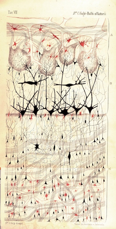
A rubbery lump, the human brain swirling in a specimen jar is an unimposing sight—more an overgrown mushroom than the seat of consciousness. The old gray matter is just that: gray. But when depicted by skilled anatomists or subjected to microscopes, MRIs, and electroencephalographs by neuroscientists, the brain and its parts can offer up visually bracing displays that call to mind an array of painters—from Motherwell and Kline to Julie Mehretu and Fred Tomaselli. Portraits of the Mind begins with a sketch of the nervous system done in eleventh-century Cairo that ably represents the movement of information from eye to optic nerve to brain. Far more exacting, though, is the 1875 drawing by Camillo Golgi of a dog’s olfactory bulb (above); the Italian pathologist developed the first workable method of staining nervous tissue and thus freshening the dun-colored mass with hue. The purposefully tangled skein of cells Golgi revealed has been further articulated as technology has permitted scientists to pursue the brain down to its cells and synapses, producing images—some look like sunspots, others bread mold—so detailed that their very relation to the flesh we animate feels questionable. A recent MRI of a damaged human thalamus depicts a detonation of bright white string; the image, coldly chaotic yet alluring, presents a piece of ourselves for inspection. The accompanying text barely convinces us that this is, in fact, a freeze-frame of an assault on our ability to hear a Bartók string quartet, taste fried chicken, or feel a lover’s caress.


